An ancient Egyptian surgical text first documented the brain's surface, meninges, and cerebrospinal fluid, linking head injuries to neurological symptoms — the earliest known record of the brain as the seat of cognition. Source — Wikipedia:
The Edwin Smith papyrus, the world's oldest surviving surgical document. Written in hieratic script in ancient Egypt around 1600 B.C., the text describes anatomical observations and the examination, diagnosis, treatment, and prognosis of 48 types of medical problems in exquisite detail. Among the treatments described are closing wounds with sutures, preventing and curing infection with honey and moldy bread, stopping bleeding with raw meat, and immobilization of head and spinal cord injuries. Translated in 1930, the document reveals the sophistication and practicality of ancient Egyptian medicine. Recto Column 6 (right) and 7 (left) of the papyrus, pictured here, discuss facial trauma. (Cases 12-20) By Jeff Dahl - Edited version of Image:EdSmPaPlateVIandVIIPrintsx.jpg, Public domain, https://commons.wikimedia.org/w/index.php?curid=2854143
600 BCE
Ayurvedic texts detail nervous system concepts
Classical Ayurvedic texts including the Sushruta Samhita described the brain as the center of consciousness and identified nerves (dhamanis and siras) conducting sensory and motor signals throughout the body. Source — Wikipedia:
Nepal, Text- 12th-13th century; Images- 18th-19th century Books Ink and opaque watercolor on palm leaf Gift of Emeritus Professor and Mrs. Thomas O. Ballinger (M.87.271a-g) South and Southeast Asian Art By Unknown author - Image: http://collections.lacma.org/sites/default/files/remote_images/piction/ma-31972784-O3.jpg Gallery: http://collections.lacma.org/node/170472 archive copy at the Wayback Machine, Public domain, https://commons.wikimedia.org/w/index.php?curid=32452002
400 BCE
Hippocratic school localizes mind to brain
Hippocratic corpus authors argued the brain is the seat of intelligence, sensation, and emotion, challenging prevailing views that the heart governed mental life. Source — Wikipedia:
Bust of Hippocrates By Unidentified engraver - 1881 Young Persons' Cyclopedia of Persons and Places Upload by RedWolf 05:45, Jan 10, 2005 (UTC), Public domain, https://commons.wikimedia.org/w/index.php?curid=164808
300 BCE
Herophilus distinguishes nerves and brain structures
Herophilus of Alexandria conducted systematic dissections distinguishing sensory from motor nerves and identifying the ventricles of the brain, advancing anatomical understanding of the nervous system. Source — Wikipedia:
Zaragoza - Antigua Facultad de Medicina - Medallón - Herófilo By Ecelan - Own work, CC BY-SA 3.0 es, https://commons.wikimedia.org/w/index.php?curid=16641053
1020 CE
Ibn al-Haytham's Book of Optics on vision
Ibn al-Haytham (Alhazen) demonstrated that vision occurs via light entering the eye, not rays emitted from it, and described the eye's anatomy and visual perception processes in his seven-volume Book of Optics. Source — Wikipedia:
This picture is of the cover page of Ibn al-Haytham's "Book of Optics", 1572. By Ibn al-Haytham, Vitello, Friedrich Risner - University of Oklahoma History of Science Collections et BnF Gallica : http://gallica.bnf.fr/ark:/12148/bpt6k312873d.r=Haytham?rk=407727;2, Public domain, https://commons.wikimedia.org/w/index.php?curid=48189707
1025 CE
Ibn Sina's Canon of Medicine on senses and brain
Ibn Sina (Avicenna) systematized knowledge of the brain, cranial nerves, and sensory perception in his Canon of Medicine, and described psychiatric conditions with early neuro-psychological explanations. Source — Wikipedia:
The Persian manuscript copy of The Canon of Medicine in Museum and Mausoleum of Avicenna, Hamedan, Iran. By Coffeetalkh - Own work, CC BY-SA 3.0, https://commons.wikimedia.org/w/index.php?curid=11906621
1030 CE
Al-Razi and al-Balkhi pioneer mental health care
Al-Razi (Rhazes) and Abu Zayd al-Balkhi independently described anxiety, depression, and obsessive-compulsive phenomena, advocating psychotherapeutic and environmental treatments — early clinical psychology and neuropsychiatry. Source — Wikipedia:
1543 CE
Vesalius publishes detailed brain anatomy
Andreas Vesalius in De humani corporis fabrica provided the most accurate anatomical illustrations of the human brain and nervous system to date, correcting Galenic errors and establishing modern neuroanatomy. Source — Wikipedia:
Vesalius publishes detailed brain anatomy By Unknown author, Public domain, https://commons.wikimedia.org/w/index.php?curid=457251
1641 CE
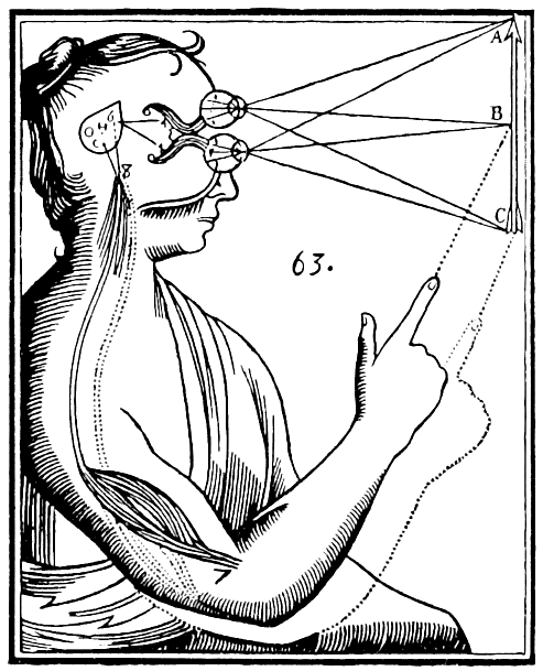
Descartes proposes mind-body interaction theory
René Descartes argued the mind and body interact through the pineal gland, framing the mind-body problem that would drive centuries of debate about consciousness and brain function. Source — Wikipedia:
Drawing from René Descartes' (1596-1650) in "Treatise of Man" supposing the function of the pineal gland. By René Descartes - Stephen Gaukroger, ed. (1998) Descartes: The World and Other Writings, Cambridge: Cambridge University Press DOI: 10.1017/CBO9780511605727. ISBN: 9780511605727., Public domain, https://commons.wikimedia.org/w/index.php?curid=1918592
1791 CE
Galvani discovers bioelectricity in nerves
Luigi Galvani showed that electrical stimulation caused frog muscle contraction, demonstrating that the nervous system operates via electrical signals — the foundation of electrophysiology. Source — Wikipedia:
Portrait of Luigi Galvani (1737-1798), Italian physicist By Unidentified painter - http://www.museopalazzopoggi.unibo.it//poggi_eng/palazzo/foto/prot http://biografieonline.it/foto-di.htm?n=Luigi+Galvani, Public domain, https://commons.wikimedia.org/w/index.php?curid=2707375Luigi Galvani's experiment 🐸⚡
1811 CE
Bell and Magendie separate sensory and motor nerves
Charles Bell and François Magendie independently established that dorsal spinal nerve roots carry sensory signals and ventral roots carry motor signals, a fundamental principle of nervous system organization. Source — Wikipedia:
Spinal cord – section By User:Polarlys - Own work, CC BY 2.5, https://commons.wikimedia.org/w/index.php?curid=1476281
1861 CE
Broca localizes speech production to frontal lobe
Paul Broca linked damage to the left frontal lobe to loss of articulate speech, providing the first strong evidence that specific cognitive functions are localized to specific brain regions. Source — Wikipedia:
Paul Broca By Unknown author - Битнер В.В. На рубеже столетий: Обзор гл. науч. и культур. приобретений XIX века. 1901. URL:http://dlib.rsl.ru/view.php?path=/rsl01003000000/rsl01003963000/rsl01003963893/rsl01003963893.pdf#?page=451, Public domain, https://commons.wikimedia.org/w/index.php?curid=16545674
1870 CE
Fritsch and Hitzig map motor cortex electrically
Gustav Fritsch and Eduard Hitzig showed that electrical stimulation of specific cortical areas in dogs produces movement, experimentally demonstrating cortical localization of motor function. Source — Wikipedia:
Gustav Fritsch By Rudolf Dührkoop - [1], Public domain, https://commons.wikimedia.org/w/index.php?curid=57525502
1873 CE
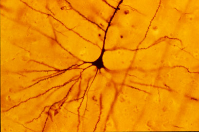
Golgi staining method reveals individual neurons
Camillo Golgi developed the silver chromate staining method that for the first time allowed visualization of individual neurons in their entirety, revolutionizing neuroanatomical research. Source — Wikipedia:
Photo by Bob Jacobs, Laboratory of Quantitative Neuromorphology Department of Psychology Colorado College https://www.coloradocollege.edu/basics/contact/directory/people/jacobs_bob.html By The original uploader was Cahass at English Wikipedia. - Transferred from en.wikipedia to Commons., CC BY-SA 3.0, https://commons.wikimedia.org/w/index.php?curid=651365
1889 CE
Cajal establishes neuron doctrine
Santiago Ramón y Cajal used Golgi staining to demonstrate that the nervous system is composed of discrete individual cells rather than a continuous network, establishing the neuron doctrine. Source — Wikipedia:
Drawing of the cells of the chick cerebellum by Santiago Ramón y Cajal, from "Estructura de los centros nerviosos de las aves", Madrid, 1905?. By Santiago Ramón - http://www.sharpbrains.com/wp-content/uploads/2008/02/cajal-chick-cerebellum.jpg, Public domain, https://commons.wikimedia.org/w/index.php?curid=555397How did Santiago Ramon y Cajal discover the neuron doctrine?
1897 CE
Sherrington coins the term 'synapse'
Charles Scott Sherrington introduced the word 'synapse' to describe the junction between neurons, conceptualizing how signals pass between nerve cells and laying groundwork for synaptic physiology. Source — Wikipedia:
Charles Scott Sherrington By Unknown author - https://wellcomeimages.org/indexplus/image/L0014994.html, CC BY 4.0, https://commons.wikimedia.org/w/index.php?curid=33623905
1906 CE
Nobel Prize honors Golgi and Cajal
The Nobel Prize in Physiology or Medicine was awarded jointly to Camillo Golgi and Santiago Ramón y Cajal for their work on the structure of the nervous system, though they held opposing views on its organization. · Wikipedia: https://en.wikipedia.org/wiki/Santiago_Ram%C3%B3n_y_Cajal
Santiago Ramón y Cajal. Spanish Nobel laureate in medicine. Restored jpg version of File:Cajal.PNG. Original image was a 5 Mb png but restoration was performed in tiff. File was mildly cropped. Dust and scratches were removed and levels were adjusted.Finally it was changed to jpg. By Original photo is anonymous although published by Clark University in 1899. Restoration by Garrondo - Cajal.PNG, Public domain, https://commons.wikimedia.org/w/index.php?curid=12334552
1921 CE
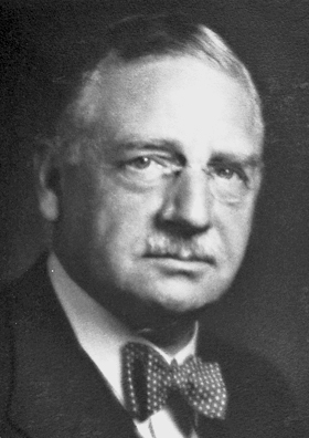
Loewi proves chemical synaptic transmission
Otto Loewi demonstrated that nerve impulses are transmitted chemically by collecting fluid from a stimulated frog heart and showing it slowed a second heart, proving chemical neurotransmission. Source — Wikipedia:
Otto Loewi, Nobel Prize in Physiology or Medicine 1936 By Albert Hilscher - https://www.nobelprize.org/prizes/medicine/1936/loewi/facts/, Public domain, https://commons.wikimedia.org/w/index.php?curid=18336306
1924 CE
Berger records first human EEG
Hans Berger recorded the first human electroencephalogram, measuring electrical activity of the brain through the scalp and discovering alpha and beta brain waves — the birth of neuroimaging. Source — Wikipedia:
HansBerger_Univ_Jena.jpeg Photo of Hans Berger (1873-1941), inventor of the electroencephalogram (EEG) and Rector of Friedrich Schiller University of Jena (1927-1938) By Unknown author - http://www.psychiatrie.uk-j.de/Geschichte.html, Public domain, https://commons.wikimedia.org/w/index.php?curid=12160449
1949 CE
Hebb proposes learning through synaptic strengthening
Donald Hebb proposed that neurons that fire together wire together, formulating the principle of synaptic plasticity as the basis for learning and memory — now known as Hebbian learning. Source — Wikipedia:
1952 CE
Hodgkin and Huxley model action potential
Alan Hodgkin and Andrew Huxley used voltage clamp experiments on squid giant axons to describe how ion fluxes generate action potentials, providing the first quantitative model of nerve impulse propagation. Source — Wikipedia:
Schematic of the basic Hodgkin-Huxley model of a neuron. By Krishnavedala - Own work, CC0, https://commons.wikimedia.org/w/index.php?curid=21725464Hodgkin and Huxley model for Action Potential Simulation
1962 CE
Hubel and Wiesel discover cortical feature detectors
David Hubel and Torsten Wiesel discovered that neurons in the visual cortex respond to specific features like edges and orientation, revealing hierarchical processing in the brain. #contemporary #science
1973 CE
Bliss and Lømo discover long-term potentiation
Timothy Bliss and Terje Lømo demonstrated that brief high-frequency stimulation of hippocampal pathways produces lasting increases in synaptic strength, defining LTP as a key mechanism of synaptic plasticity. Source — Wikipedia:
An example of long-term potentiation recorded at Schaffer collateral/CA1 synapses in the hippocampus of rat brain. Graph: The rising slope of an extracellular EPSP field recorded in stratum radiatum of area CA1 in response to an extracellular stimulation of Schaffer collaterals. Test stimulus is a single shock of 100 us duration, approximately .01 mA. Tetanus is the test stimulus delivered at 100 Hz for 1s. Inset: a diagram of a hippocampal slice showing the electrode placement. Above the diagram are examples of the raw EPSP field potentials before and after tetanus. Extended caption: An example of long-term potentiation (LTP). The graph illustrates the analysis of a single field potential recording from a rat hippocampal slice. The inset shows the placement of the stimulating and recording electrode within the slice, and above, two raw traces of EPSP field potentials before and after tetanic stimulation. Field potentials were evoked by stimulation of Schaffer collaterals and recorded in stratum radiatum of the hippocampal slice (i.e. the Schaffer collateral/CA1 synapses). The individual points on the graph each represent the measurement of the rising slope of one EPSP field potential. Black squares indicate the measurements taken before tetanic stimulation. The green squares are measurements taken immediately after tetanic stimulation (PTP or post-tetanic potentiation) while the blue squares are measurements taken between 3 and 60 minutes after tetanization (LTP or long-term potentiation). Test stimuli were administered once every 30 sec. Tetanic stimulation was the test stimulus given at 100 Hz for 1 sec. Note how the amplitude of the EPSP field settles into a new, more elevated level after tetanic stimulation. By Synaptidude at English Wikipedia - This image created by Synaptidude 23:45, 15 August 2005 (UTC) , assembled in CorelDraw!, CC BY-SA 3.0, https://commons.wikimedia.org/w/index.php?curid=886855
1977 CE
PET scanning enables functional brain imaging
Positron emission tomography was developed for human brain imaging, allowing researchers to visualize metabolic activity across brain regions during cognitive tasks — a major advance in functional neuroimaging. Source — Wikipedia:
Positron emission tomography machine By Spacecoastcreative - Own work, CC0, https://commons.wikimedia.org/w/index.php?curid=149063982
1986 CE
Kandel links synaptic plasticity to memory molecules
Eric Kandel showed that short- and long-term memory in Aplysia involve distinct molecular mechanisms of synaptic plasticity, bridging synaptic physiology with molecular biology of learning. Source — Wikipedia:
Eric Kandel By Bengt Oberger - Own work, CC BY-SA 4.0, https://commons.wikimedia.org/w/index.php?curid=37241660
1990 CE
fMRI revolutionizes functional brain imaging
Seiji Ogawa and colleagues demonstrated that blood oxygenation level-dependent (BOLD) signals could map brain activity noninvasively using MRI, launching the era of fMRI cognitive neuroscience. Source — Wikipedia:
Figure 12.7 fMRI in Version 8.25 from the Textbook OpenStax Anatomy and Physiology Published May 18, 2016 Access for free at https://openstax.org/books/anatomy-and-physiology/pages/1-introduction By OpenStax - https://openstax.org/books/anatomy-and-physiology/pages/12-1-basic-structure-and-function-of-the-nervous-system, CC BY 4.0, https://commons.wikimedia.org/w/index.php?curid=30147912Functional Magnetic Resonance Imaging (fMRI) explained | Neuroscience Methods 101
1992 CE
Decade of the Brain boosts neuroscience research
The U.S. Congress designated the 1990s as the Decade of the Brain, increasing funding and public awareness for neuroscience research across synaptic transmission, plasticity, and imaging domains. Source — Wikipedia:
Logo of the Decade of the Brain initiative. By US Gov. - https://www.loc.gov/loc/brain/, Public domain, https://commons.wikimedia.org/w/index.php?curid=37228176
2000 CE
Nobel Prize for signal transduction in the brain
Arvid Carlsson, Paul Greengard, and Eric Kandel received the Nobel Prize for discoveries concerning signal transduction in the nervous system, including dopamine as a neurotransmitter and molecular mechanisms of memory. · Wikipedia: https://en.wikipedia.org/wiki/Arvid_Carlsson
Swedish pharmacologist and Nobel laureate Arvid Carlsson giving a lecture at the 2011 Göteborg Science Festival. By Vogler - Own work, CC BY-SA 3.0, https://commons.wikimedia.org/w/index.php?curid=154865850
2013 CE
BRAIN Initiative launched
The U.S. BRAIN Initiative was announced to accelerate the development and application of neurotechnologies for understanding brain circuit function, spurring global brain research efforts. Source — Wikipedia:
President Obama Speaks on the BRAIN Initiative and American Innovation
2023 CE
Whole mouse brain connectome mapped
Researchers published the most complete connectome of a mouse brain, mapping over 200,000 neurons and 500 million synapses, providing an unprecedented resource for understanding brain circuit organization. Source — Wikipedia:
diffusion MRI Tractography in the brain white matter. By Xavier Gigandet et. al. - Gigandet X, Hagmann P, Kurant M, Cammoun L, Meuli R, et al. (2008) Estimating the Confidence Level of White Matter Connections Obtained with MRI Tractography. PLoS ONE 3(12): e4006. doi:10.1371/journal.pone.0004006, CC BY 2.5, https://commons.wikimedia.org/w/index.php?curid=8134159