Encyclopedia/1. The Cosmos & The Natural World/5. Life Sciences/05. Organismal Biology • Curated by Admin Timeline.sg
Embryology · Morphogenesis
Chronological Storyline (29 Milestones)
600 BCE
Sushruta Samhita describes surgery and anatomy
The Sushruta Samhita, an ancient Indian medical text attributed to Sushruta, documented surgical techniques, human anatomy, and developmental physiology, establishing foundations for systematic medical practice in India. Source — Wikipedia:
Nepal, Text- 12th-13th century; Images- 18th-19th century Books Ink and opaque watercolor on palm leaf Gift of Emeritus Professor and Mrs. Thomas O. Ballinger (M.87.271a-g) South and Southeast Asian Art By Unknown author - Image: http://collections.lacma.org/sites/default/files/remote_images/piction/ma-31972784-O3.jpg Gallery: http://collections.lacma.org/node/170472 archive copy at the Wayback Machine, Public domain, https://commons.wikimedia.org/w/index.php?curid=32452002
300 BCE
Aristotle observes embryonic development
Aristotle described the development of chick embryos and proposed epigenesis — the idea that form arises gradually from unformed matter — foundational to embryology for two millennia. Source — Wikipedia:
it:Octopus macropus By SUBnormali Team Original uploader was Yoruno at it.wikipedia - Transferred from it.wikipedia (Original text : subnormali-team.blogspot.com), CC BY-SA 3.0, https://commons.wikimedia.org/w/index.php?curid=6381781
200 BCE
Huangdi Neijing compiled
The Huangdi Neijing, the foundational text of Chinese medicine, described physiological development, organ systems, and the circulation of qi, influencing East Asian medical thought for over two thousand years. Source — Wikipedia:
Huangdi Neijing (黄帝内経, kōteidaikei), Myōdō (明堂) By Unknown author - [1], Public domain, https://commons.wikimedia.org/w/index.php?curid=11662446
165 CE
Galen writes on embryology and anatomy
Galen produced extensive anatomical and physiological writings including "De Formatione Foetuum," describing embryonic organ formation and the three souls concept, dominating Western medicine for centuries. Source — Wikipedia:
Portrait of Galen. By Georg Paul Busch (engraver) - The Lancet, Public domain, https://commons.wikimedia.org/w/index.php?curid=40855568
1025 CE
Ibn Sina completes the Canon of Medicine
Ibn Sina (Avicenna) completed al-Qanun fi al-Tibb (Canon of Medicine), synthesizing Greco-Islamic medical knowledge including embryology, anatomy, and physiology, becoming the standard medical text in Europe for centuries. Source — Wikipedia:
The Persian manuscript copy of The Canon of Medicine in Museum and Mausoleum of Avicenna, Hamedan, Iran. By Coffeetalkh - Own work, CC BY-SA 3.0, https://commons.wikimedia.org/w/index.php?curid=11906621
1242 CE
Ibn al-Nafis discovers pulmonary circulation
Ibn al-Nafis described the pulmonary circulation of blood, correcting Galen's anatomy and providing a physiological framework essential to understanding developmental and adult cardiovascular systems. Source — Wikipedia:
Photograph of statue of Ibn Al Nafis By Unknown author - Arabic book on ibn al Nafis Takrouri M.S. M & Khalaf M 2003, CC BY-SA 3.0, https://commons.wikimedia.org/w/index.php?curid=4759221
1500 CE
Leonardo da Vinci draws human embryos
Leonardo da Vinci produced detailed anatomical drawings of the human fetus in utero, depicting its position and the placenta with unprecedented accuracy for his time. Source — Wikipedia:
Leonardo da Vinci draws human embryos By Attributed to Francesco Melzi - File:Francesco_Molzi_-_Portrait_of_Leonardo_-_WGA14795.jpg, Public domain, https://commons.wikimedia.org/w/index.php?curid=78645105Leonardo da Vinci The Fetus and Lining of the Uterus
1651 CE
Harvey publishes on generation
William Harvey published "Exercitationes de Generatione Animalium," arguing for epigenesis and that all animals arise from eggs, building on his discovery of blood circulation. Source — Wikipedia:
Harvey publishes on generation By After Daniël Mijtens - National Portrait Gallery: NPG 5115, Public domain, https://commons.wikimedia.org/w/index.php?curid=5975113
1672 CE
de Graaf describes ovarian follicles
Regnier de Graaf described the ovarian structures later named Graafian follicles, advancing understanding of female reproductive anatomy and its role in generation. Source — Wikipedia:
portret van Reinier de Graaf By Unknown author - Museum Boijmans Van Beuningen, Public domain, https://commons.wikimedia.org/w/index.php?curid=2891214
1675 CE
Leeuwenhoek observes spermatozoa
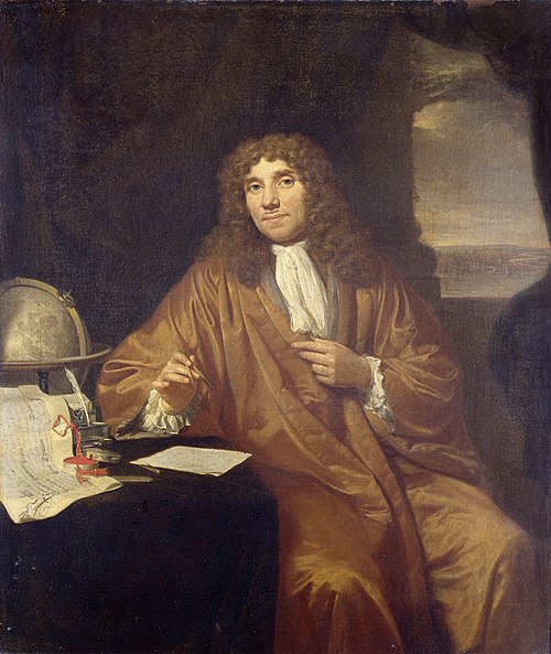
Antonie van Leeuwenhoek used his microscope to observe spermatozoa, sparking the preformationist debate over whether the embryo was preformed in the sperm or the egg. Source — Wikipedia:
Portrait of Anthonie van Leeuwenhoek (1632-1723), natural philosopher, in Delft. Knee piece, sitting at a writing table on which a certificate lies from his appointment as a member of the Royal Society in London by Charles II. Also a globe and an ink set; He holds a compass in his hand. By Jan Verkolje - http://www.rijksmuseum.nl/collectie/SK-A-957, Public domain, https://commons.wikimedia.org/w/index.php?curid=34320547
1759 CE
Wolff proposes epigenesis in development
Caspar Friedrich Wolff argued in "Theoria Generationis" that embryonic tissues develop progressively from undifferentiated layers, reviving epigenesis against preformationism. Source — Wikipedia:
Caspar Friedrich Wolff, German physician, paper cut, before 1770 By F. Anting Liftarn - Based on Image:Caspar Friedrich Wolff.jpg, Public domain, https://commons.wikimedia.org/w/index.php?curid=1115580
1828 CE
von Baer discovers the mammalian ovum
Karl Ernst von Baer discovered the mammalian ovum and established the germ layer theory, founding modern comparative embryology and its relation to evolutionary thought. Source — Wikipedia:
Illustrasjon hentet fra boken "Voyages de la Commission scientifique du Nord, en Scandinavie, en Laponie, au Spitzberg et aux Feröe" av ukjent forfatter og utgitt av Arthus BertrandParis : Arthus Bertrand, [1852] (Paris, [1852]) By Unknown author - National Library of Norway, Public domain, https://commons.wikimedia.org/w/index.php?curid=34041566
1859 CE
Darwin links embryology to evolution
Charles Darwin used embryological evidence in "On the Origin of Species" to support common descent, noting that embryos of related species resemble each other more than adults do. Source — Wikipedia:
Cropped and rotated version of File:Origin of Species.jpg, which is of the title page and the facing page of the 1859 Murray edition of the Origin of species by Charles Darwin. By John Murray, Publisher - This image has an extracted file: Origin of Species.jpg., Public domain, https://commons.wikimedia.org/w/index.php?curid=320416The Making of a Theory: Darwin, Wallace, and Natural Selection — HHMI BioInteractive Video
1866 CE
Haeckel formulates the biogenetic law
Ernst Haeckel proposed that ontogeny recapitulates phylogeny — that embryonic development replays evolutionary history — influencing developmental biology despite later criticism. Source — Wikipedia:
1888 CE
Driesch demonstrates developmental regulation
Hans Driesch separated sea urchin blastomeres and showed each could develop into a complete larva, demonstrating regulative development and challenging preformationist ideas. Source — Wikipedia:
Hans Driesch, a philosopher of science By Unknown author - www.merke.ch, Public domain, https://commons.wikimedia.org/w/index.php?curid=15121784
1891 CE
Hans Spemann begins morphogenesis experiments
Hans Spemann began his pioneering work on embryonic induction and morphogenesis, later using hair-loop constriction experiments on salamander embryos to study cell fate determination. Source — Wikipedia:
Hans Spemann (June 27, 1869 – September 9, 1941) By Unknown author - http://www.nobelprize.org/nobel_prizes/medicine/laureates/1935, Public domain, https://commons.wikimedia.org/w/index.php?curid=18336155
1924 CE
Spemann and Mangold discover the organizer
Hilde Mangold and Hans Spemann demonstrated that the dorsal lip of the blastopore — the organizer — induces secondary body axis formation in amphibian embryos, a landmark in morphogenesis. Source — Wikipedia:
Hilde Mangold and the Organizer of Life | Great Minds
1942 CE
Schmalhausen publishes on stabilizing selection
Ivan Schmalhausen published "Factors of Evolution," integrating developmental processes with evolutionary theory and contributing to the foundations of evolutionary developmental biology. Source — Wikipedia:
1951 CE
Turing proposes morphogen theory
Alan Turing proposed that chemical substances called morphogens reacting and diffusing could generate pattern formation in development, founding the mathematical theory of morphogenesis. · Wikipedia: https://en.wikipedia.org/wiki/Morphogenesis
1952 CE
Briggs and King perform nuclear transfer
Robert Briggs and Thomas King transplanted nuclei from frog blastula cells into enucleated eggs, producing normal tadpoles and founding nuclear transplantation in developmental biology. Source — Wikipedia:
1961 CE
Nieuwkoop discovers mesoderm induction
Pieter Nieuwkoop demonstrated that vegetal blastomeres induce overlying equatorial cells to become mesoderm in amphibian embryos, establishing the concept of mesoderm induction in development. #contemporary #science
1984 CE
Homeobox genes discovered in vertebrates
The discovery that homeobox (Hox) genes are conserved between Drosophila and vertebrates revealed that the genetic toolkit patterning the body plan is shared across the animal kingdom. · Wikipedia: https://en.wikipedia.org/wiki/Homeobox
The Antennapedia homeodomain protein from Drosophila melanogaster bound to a DNA fragment, illustrating the binding interactions of the recognition helix and unstructured N-terminus with the DNA major and minor grooves. By Opabinia regalis - Self created from PDB entry 1AHD using the freely available visualization and analysis package VMD, CC BY-SA 3.0, https://commons.wikimedia.org/w/index.php?curid=1024362
1988 CE
Nüsslein-Volhard identifies morphogen gradients
Christiane Nüsslein-Volhard and Eric Wieschaus's systematic mutagenesis screen identified key genes controlling embryonic patterning in Drosophila, earning them the Nobel Prize and revealing conserved morphogen pathways. Source — Wikipedia:
Christiane Nüsslein-Volhard By Rama - Own work, CC BY-SA 2.0 fr, https://commons.wikimedia.org/w/index.php?curid=2270623
1995 CE
Nobel Prize for embryonic patterning genes
The Nobel Prize in Physiology or Medicine was awarded to Edward Lewis, Christiane Nüsslein-Volhard, and Eric Wieschaus for discoveries of genetic control of early embryonic development. · Wikipedia: https://en.wikipedia.org/wiki/Nobel_Prize_in_Physiology_or_Medicine
Description: Front side (obverse) of one of the Nobel Prize medals in Physiology or Medicine awarded in 1950 to researchers at the Mayo Clinic in Rochester, Minnesota. By Photograph: JonathunderMedal: Erik Lindberg (1873-1966) - Derivative of File:NobelPrize.JPG, PD-US, https://en.wikipedia.org/w/index.php?curid=58432969
1996 CE
Dolly the sheep cloned by nuclear transfer
Ian Wilmut and Keith Campbell at the Roslin Institute cloned Dolly the sheep from an adult somatic cell nucleus, demonstrating that differentiated cells can be reprogrammed to support full development. · Wikipedia: https://en.wikipedia.org/wiki/Dolly_(sheep)
The Story of Dolly the Cloned Sheep | Retro Report | The New York Times
1998 CE
Human embryonic stem cells isolated
James Thomson and colleagues isolated and cultured human embryonic stem cells, opening new avenues for studying human development and regenerative medicine. · Wikipedia: https://en.wikipedia.org/wiki/Stem_cell
Transmission electron micrograph of a mesenchymal stem cell displaying typical ultrastructural characteristics. By Robert M. Hunt - Own work, CC BY 3.0, https://commons.wikimedia.org/w/index.php?curid=28384153
2006 CE
Yamanaka creates induced pluripotent stem cells
Shinya Yamanaka reprogrammed mouse fibroblasts to pluripotent stem cells using four transcription factors, revolutionizing developmental biology and earning the Nobel Prize in 2012. · Wikipedia: https://en.wikipedia.org/wiki/Induced_pluripotent_stem_cell
Researchers at NIH's National Eye Institute (NEI) developed the first patient-derived stem cell model for studying eye conditions related to oculocutaneous albinism (OCA). Learn more: at https://www.nih.gov/news-events/news-releases/nih-researchers-develop-first-stem-cell-model-albinism-study-related-eye-conditions In this image: A human induced pluripotent stem cell colony from OCA1A patient. The image was acquired using a confocal microscope and is stained for pluripotency marker proteins. The red color depicts transcription factor OCT4, green is SSEA4 protein and blue represents the nucleus of the cells. Credit: National Eye Institute/NIH By NIH Image Gallery from Bethesda, Maryland, USA - Human induced pluripotent stem cell colony, Public domain, https://commons.wikimedia.org/w/index.php?curid=114900547Scientist Stories: Shinya Yamanaka, Cell reprogramming and Pioneering Induced Pluripotent Stem Cells
2010 CE
Optogenetics applied to developmental biology
Optogenetic tools were adapted to control gene expression and signaling with light in living embryos, enabling precise spatiotemporal manipulation of developmental processes. Source — Wikipedia:
2022 CE
Synthetic embryos grown from stem cells
Researchers grew mouse embryo models from stem cells without sperm or egg, reaching advanced developmental stages with beating hearts and brain folds, advancing synthetic embryology. · Wikipedia: https://en.wikipedia.org/wiki/Synthetic_embryo



![Illustrasjon hentet fra boken "Voyages de la Commission scientifique du Nord, en Scandinavie, en Laponie, au Spitzberg et aux Feröe" av ukjent forfatter og utgitt av Arthus BertrandParis : Arthus Bertrand, [1852] (Paris, [1852])](https://upload.wikimedia.org/wikipedia/commons/thumb/7/7a/Voyages_de_la_Commission_scientifique_du_Nord%2C_en_Scandinavie%2C_en_Laponie%2C_au_Spitzberg_et_aux_Fer%C3%B6e_-_no-nb_digibok_2009040211001-118.jpg/500px-Voyages_de_la_Commission_scientifique_du_Nord%2C_en_Scandinavie%2C_en_Laponie%2C_au_Spitzberg_et_aux_Fer%C3%B6e_-_no-nb_digibok_2009040211001-118.jpg)