Encyclopedia/1. The Cosmos & The Natural World/5. Life Sciences/03. Molecular & Cellular Biology • Curated by Admin Timeline.sg
Signal transduction · Membrane biology
Chronological Storyline (30 Milestones)
300 BCE
Charaka and Sushruta record physiology
Ancient Indian medical texts by Charaka and Sushruta described body functions, tissues, and channels for vital fluids, representing early physiological thought in the Indian tradition. Source — Wikipedia:
Nepal, Text- 12th-13th century; Images- 18th-19th century Books Ink and opaque watercolor on palm leaf Gift of Emeritus Professor and Mrs. Thomas O. Ballinger (M.87.271a-g) South and Southeast Asian Art By Unknown author - Image: http://collections.lacma.org/sites/default/files/remote_images/piction/ma-31972784-O3.jpg Gallery: http://collections.lacma.org/node/170472 archive copy at the Wayback Machine, Public domain, https://commons.wikimedia.org/w/index.php?curid=32452002
280 BCE
Herophilus and Erasistratus dissect bodies
In Hellenistic Alexandria, Herophilus and Erasistratus performed systematic human dissections, distinguishing nerves from blood vessels and studying organ function, laying early foundations for physiology. Source — Wikipedia:
Zaragoza - Antigua Facultad de Medicina - Medallón - Herófilo By Ecelan - Own work, CC BY-SA 3.0 es, https://commons.wikimedia.org/w/index.php?curid=16641053
1021 CE
Ibn al-Haytham writes on vision
Ibn al-Haytham's Book of Optics described light entering the eye and laid experimental groundwork for understanding sensory physiology, influencing later European science. Source — Wikipedia:
Cropped version of the frontispiece of Johannes Hevelius, Selenographia, depicting Ibn al-Haytham (Alhazen) By Adolph Boÿ, engraved by Jeremias Falck. Used as the frontispiece to Johannes Hevelius, Selenographia, 1647 - https://www.loc.gov/resource/rbctos.2017rosen1321/?sp=9&r=0.059,0.617,0.327,0.182,0, Public domain, https://commons.wikimedia.org/w/index.php?curid=165125519
1241 CE
Ibn al-Nafis describes pulmonary circulation
In Cairo, Ibn al-Nafis described the circulation of blood through the lungs, correcting Galenic physiology centuries before William Harvey. Source — Wikipedia:
Photograph of statue of Ibn Al Nafis By Unknown author - Arabic book on ibn al Nafis Takrouri M.S. M & Khalaf M 2003, CC BY-SA 3.0, https://commons.wikimedia.org/w/index.php?curid=4759221
1651 CE
Harvey publishes on blood circulation
William Harvey demonstrated that blood circulates continuously through the body pumped by the heart, establishing experimental physiology as a discipline. Source — Wikipedia:
Harvey publishes on blood circulation By After Daniël Mijtens - National Portrait Gallery: NPG 5115, Public domain, https://commons.wikimedia.org/w/index.php?curid=5975113Misunderstood Geniuses: William Harvey | World Science Festival
1665 CE
Hooke coins the word 'cell'
Robert Hooke observed cork under a microscope and coined the term 'cell' in Micrographia, marking the first description of cellular structure. Source — Wikipedia:
Title page By Robert Hooke (1635-1703) - This image is available from the National Library of Wales, Public domain, https://commons.wikimedia.org/w/index.php?curid=7442137Robert Hook coined the word Cell | BBC
1674 CE
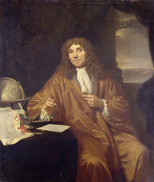
Leeuwenhoek observes living cells
Antonie van Leeuwenhoek used a single-lens microscope to observe protozoa and bacteria, providing the first glimpses of living single-celled organisms. Source — Wikipedia:
Portrait of Anthonie van Leeuwenhoek (1632-1723), natural philosopher, in Delft. Knee piece, sitting at a writing table on which a certificate lies from his appointment as a member of the Royal Society in London by Charles II. Also a globe and an ink set; He holds a compass in his hand. By Jan Verkolje - http://www.rijksmuseum.nl/collectie/SK-A-957, Public domain, https://commons.wikimedia.org/w/index.php?curid=34320547
1838 CE
Cell theory proposed by Schleiden and Schwann
Matthias Schleiden and Theodor Schwann proposed that all living organisms are composed of cells, establishing cell theory as a unifying biological principle. Source — Wikipedia:
HeLa cells stained with Hoechst 33258 stain. By TenOfAllTrades at English Wikipedia - Own work, Public domain, https://commons.wikimedia.org/w/index.php?curid=1583880
1855 CE
Virchow: cells arise from cells
Rudolf Virchow stated that all cells arise from pre-existing cells, completing the classical cell theory and founding modern cellular pathology. Source — Wikipedia:
Rudolf Virchow By Unknown author - http://ihm.nlm.nih.gov/images/B25666, Public domain, https://commons.wikimedia.org/w/index.php?curid=19353800
1869 CE
Miescher discovers nuclein (DNA)
Friedrich Miescher isolated nuclein from white blood cells, discovering what would later be identified as DNA, the molecule of heredity. Source — Wikipedia:
Friedrich Miescher (scientist) By Unknown author - copied from http://www.pbs.org/wgbh/nova/photo51/images/befo-miescher.jpg, Public domain, https://commons.wikimedia.org/w/index.php?curid=789048
1890 CE
Overton discovers membrane lipid nature
Charles Overton showed that substances soluble in lipids enter cells more readily, leading to the inference that the cell membrane is lipid in nature. Source — Wikipedia:
The cell membrane, also called the plasma membrane or plasmalemma, is a semipermeable lipid bilayer common to all living cells. It contains a variety of biological molecules, primarily proteins and lipids, which are involved in a vast array of cellular processes. It also serves as the attachment point for both the intracellular cytoskeleton and, if present, the cell wall. By derivative work: Dhatfield (talk) Cell_membrane_detailed_diagram_3.svg: *derivative work: Dhatfield (talk) Cell_membrane_detailed_diagram.svg: LadyofHats Mariana Ruiz - Cell_membrane_detailed_diagram_3.svg, CC BY-SA 3.0, https://commons.wikimedia.org/w/index.php?curid=4228810
1897 CE
Buchner shows cell-free fermentation
Eduard Buchner demonstrated that yeast extracts could ferment sugar without living cells, proving that metabolism is driven by enzymes and founding biochemistry. Source — Wikipedia:
Eduard Buchner, Nobel Laureate in Chemistry (1907) By Unknown author - http://www.nobelprize.org/nobel_prizes/chemistry/laureates/1907/buchner.html, Public domain, https://commons.wikimedia.org/w/index.php?curid=18253176
1902 CE
Loeb demonstrates artificial parthenogenesis
Jacques Loeb showed that unfertilized sea urchin eggs could be induced to develop by chemical treatment, revealing that cell behavior could be controlled by external chemical signals. Source — Wikipedia:
Jacques Loeb By unknown/pseudonymous - Images from the History of Medicine (NLM) [1], Public domain, https://commons.wikimedia.org/w/index.php?curid=9006359
1925 CE
Gorter and Grendel propose lipid bilayer
Evert Gorter and François Grendel proposed that the cell membrane consists of a lipid bilayer, based on extracted lipid areas from red blood cell membranes. Source — Wikipedia:
Schem of the lipid bilayer on viruses, i.e. By Bensaccount - http://en.wikipedia.org/wiki/Image:Lipid_bilayer_section.gif, Public domain, https://commons.wikimedia.org/w/index.php?curid=2929946
1935 CE
Danielli and Davson model the membrane
Hugh Davson and James Danielli proposed the sandwich model of the cell membrane with a lipid bilayer coated by proteins, the first detailed membrane model. Source — Wikipedia:
1948 CE
Hodgkin and Keynes study ion fluxes
Alan Hodgkin and Richard Keynes used radioactive isotopes to measure ion fluxes across nerve membranes, advancing understanding of the cellular basis of nerve impulses. Source — Wikipedia:
Alan Lloyd Hodgkin, Nobel Prize in Physiology or Medicine 1963 By Unknown author - https://www.nobelprize.org/nobel_prizes/medicine/laureates/1963/, Public domain, https://commons.wikimedia.org/w/index.php?curid=18346641
1952 CE
Hodgkin and Huxley model action potentials
Alan Hodgkin and Andrew Huxley recorded intracellularly from squid giant axons and formulated a mathematical model of the action potential, founding cellular electrophysiology. Source — Wikipedia:
Schematic of the basic Hodgkin-Huxley model of a neuron. By Krishnavedala - Own work, CC0, https://commons.wikimedia.org/w/index.php?curid=21725464
1957 CE
Sutherland discovers cyclic AMP
Earl Sutherland discovered cyclic AMP as a second messenger mediating the action of hormones like epinephrine, founding the field of signal transduction. Source — Wikipedia:
1970 Award Winner - Earl W. Sutherland, Jr. By Unknown author - http://resource.nlm.nih.gov/101441404, Public domain, https://commons.wikimedia.org/w/index.php?curid=71024713
1961 CE
Hayflick limit discovered
Leonard Hayflick demonstrated that normal somatic cells divide a limited number of times in culture, establishing the concept of cellular senescence. Source — Wikipedia:
1972 CE
Tu Youyou isolates artemisinin
Tu Youyou isolated artemisinin from Artemisia annua, a drug effective against malaria parasites, drawing on traditional Chinese medical texts and modern cellular pharmacology. Source — Wikipedia:
Nobel Laureates in medicin 2015 By Bengt Nyman from Vaxholm, Sweden - 810_4987 Tu Youyou, medicine, CC BY 2.0, https://commons.wikimedia.org/w/index.php?curid=123803054Tu Youyou: Discovery of Artemisinin | Heroes of Progress | Ep. 25
1972 CE
Singer and Nicolson propose fluid mosaic model
Seymour Jonathan Singer and Garth Nicolson proposed the fluid mosaic model, describing the cell membrane as a dynamic lipid bilayer with embedded proteins. Source — Wikipedia:
The cell membrane, also called the plasma membrane or plasmalemma, is a semipermeable lipid bilayer common to all living cells. It contains a variety of biological molecules, primarily proteins and lipids, which are involved in a vast array of cellular processes. It also serves as the attachment point for both the intracellular cytoskeleton and, if present, the cell wall. By Mariana Ruiz - Own work, Public domain, https://commons.wikimedia.org/w/index.php?curid=6027169
1976 CE
Neher and Sakmann develop patch clamp
Erwin Neher and Bert Sakmann developed the patch clamp technique, enabling the recording of currents through single ion channels and revolutionizing membrane biology. Source — Wikipedia:
This is a patched bacterial en:spheroplast on a glass pipette. I took this picture myself, and hereby release it to the public domain By Vlad75 at English Wikipedia - Transferred from en.wikipedia to Commons., Public domain, https://commons.wikimedia.org/w/index.php?curid=5072761
1977 CE
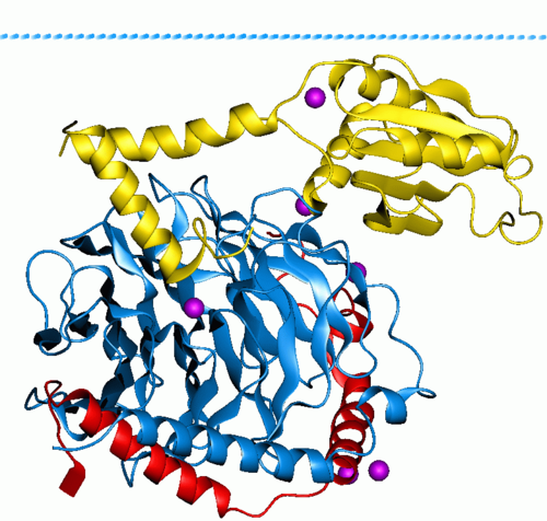
Gilman and Rodbell discover G-proteins
Martin Rodbell and Alfred Gilman discovered G-proteins that transmit signals from cell-surface receptors to intracellular effectors, a central mechanism in signal transduction. Source — Wikipedia:
Protein image from OPM database Phosducin- transducin beta-gamma complex. Beta and gamma subunits of G-protein are shown by blue and red, respectively. By Andrei Lomize - Own work, Public domain, https://commons.wikimedia.org/w/index.php?curid=34114642
1981 CE
Nobel Prize awarded for split genes
Philip Sharp and Richard Roberts discovered split genes and RNA splicing, revealing that eukaryotic gene expression involves complex cellular processing of transcripts. Source — Wikipedia:
General process of RNA splicing By Original: Iinaba Vector: Masumrezarock100 - https://commons.wikimedia.org/w/index.php?title=File:Splicesome.pdf, CC BY-SA 4.0, https://commons.wikimedia.org/w/index.php?curid=115983282
1986 CE
Berridge links IP3 to calcium signaling
Michael Berridge showed that inositol trisphosphate (IP3) releases calcium from intracellular stores, establishing a key second-messenger pathway in signal transduction. Source — Wikipedia:
1992 CE
Preston and Agre characterize aquaporin
Peter Agre and colleagues identified aquaporin-1 as the long-sought water channel protein, explaining how cells rapidly transport water across membranes. Source — Wikipedia:
Sideview of Aquaporin 1 Channel, PDB ID, 1J4N generated by PyMOL. Work of User:Vossman. By Vossman - Own work, CC BY-SA 3.0, https://commons.wikimedia.org/w/index.php?curid=1445738
1994 CE
Tsien and Chalfie develop GFP imaging
Roger Tsien, Martin Chalfie, and Osamu Shimomura developed green fluorescent protein technology, enabling real-time visualization of proteins in living cells. Source — Wikipedia:
Cartoon representation of the molecular structure of protein registered with 1ema code. By Jawahar Swaminathan and MSD staff at the European Bioinformatics Institute - https://www.ebi.ac.uk/pdbe/static/entry/1ema_deposited_chain_front_image-800x800.png, displayed on https://www.ebi.ac.uk/pdbe/entry/pdb/1ema, Public domain, https://commons.wikimedia.org/w/index.php?curid=6307286
2003 CE
MacKinnon solves potassium channel structure
Roderick MacKinnon determined the three-dimensional structure of a potassium ion channel by X-ray crystallography, revealing the molecular basis of ion selectivity. Source — Wikipedia:
Roderick MacKinnon, M.D. By PotassiumChannel - Own work, CC BY-SA 3.0, https://commons.wikimedia.org/w/index.php?curid=153692136
2012 CE
Lefkowitz and Kobilka win for GPCR structure
Robert Lefkowitz and Brian Kobilka were recognized for discovering the inner workings of G-protein-coupled receptors, the largest family of cell-surface signal transduction proteins. Source — Wikipedia:
The human beta-2 adrenergic receptor, a G protein-coupled receptor, shown in rainbow color from N-terminus (blue) to C-terminus (red) with the extracellular surface at the top of the image. The partial inverse agonist carazolol is shown as gray sticks. Rendered using PyMol from PDB ID 2RH1. By Opabinia regalis - Own work, CC BY-SA 3.0, https://commons.wikimedia.org/w/index.php?curid=38540293
2020 CE
BGI and global teams map COVID-19 cell entry
Researchers including teams at China's BGI and international collaborators mapped how SARS-CoV-2 enters human cells via ACE2 receptors, advancing cellular-level understanding of viral infection. Source — Wikipedia:
Structure of the ACE2 protein. Based on PyMOL rendering of PDB 1r42. By Emw - Own work, CC BY-SA 3.0, https://commons.wikimedia.org/w/index.php?curid=8761587Animation of SARS-CoV-2 entry into human host-cell.